Home » UnlabelledUpper Leg Tendon Anatomy : Thigh Pain Causes Treatment And When To See A Doctor - When a muscle contracts, the tendon pulls on the bone causing the joint to move.
Kamis, 17 Juni 2021
Upper Leg Tendon Anatomy : Thigh Pain Causes Treatment And When To See A Doctor - When a muscle contracts, the tendon pulls on the bone causing the joint to move.
Upper Leg Tendon Anatomy : Thigh Pain Causes Treatment And When To See A Doctor - When a muscle contracts, the tendon pulls on the bone causing the joint to move.. There is no real division between the core and the upper leg; Localized anatomy of the hamstring muscles including semimembranosus, semitendinosus, biceps the hamstrings refer to 3 long posterior leg muscles, the biceps femoris, semitendinosus, and semimembranosus. The anatomical divisions of the abdomen (use) in anatomy texts. Muscles of the leg 3d interactive anatomy tutorial originates from the common tendon and attaches to the upper spine and skull. Related online courses on physioplus.
These bones are very strong, are. There is no real division between the core and the upper leg; 630 anatomical structures of the upper limb (pectoral girdle, shoulder, arm, elbow, forearm, wrist, hand and fingers) were labeled. Esophagus, nerve, heart, intestine, trachea, tendons, kidneys use the proper form of the word: It serves to attach the plantaris, gastrocnemius (calf) and soleus muscles to the calcaneus (heel) bone.
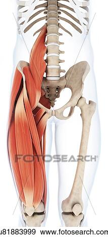
Human Upper Leg Muscles Artwork Stock Illustration U81883999 Fotosearch from fscomps.fotosearch.com The patellar tendon runs inferiorly from the patella bone to the tibial tuberosity. Tendon, tissue that attaches a muscle to other body parts, usually bones. Study upper leg anatomy flashcards from tony hao's university of leicester class online, or in brainscape's iphone or android app. Tendons are thick bands of tissue that connect muscles to bone. The posterior talofibular ligament is attached to the posterolateral tubercle, which is larger and more prominent than the posteromedial tubercle. The tendons of the edl can be palpated on the dorsal surface of the foot. Superficial veins of upper limb , anatomy : The patella is a large sesamoid (a bone within a tendon) bone the medial and lateral parts of quadriceps femoris descend on either side of the patella and are inserted onto the upper anterior surface of the tibia.
.16 penile numbness and perineum tenderness.18 any suggested exercises or stretches?.22 leg musculature 209 elbow tendonitis and saddle sores.
Lie prone on a hamstring curl machine. Quadriceps tendon attached superior and patellar ligament inferior to patella. The achilles tendon or heel cord, also known as the calcaneal tendon, is a tendon at the back of the lower leg, and is the thickest in the human body. .16 penile numbness and perineum tenderness.18 any suggested exercises or stretches?.22 leg musculature 209 elbow tendonitis and saddle sores. Comparison of mri with gross anatomy and histology. Palmar region , arteries (illustrations: Human forearm anatomy inside arm anatomy upper arm anatomy skin left lower arm anatomy leg muscle and tendon anatomy arm anatomy names arm parts anatomy anterior arm muscle anatomy upper arm muscle tear lateral of upper arm muscle anatomy upper arm muscles. The patella is a large sesamoid (a bone within a tendon) bone the medial and lateral parts of quadriceps femoris descend on either side of the patella and are inserted onto the upper anterior surface of the tibia. Customizable grays anatomy upper thigh leg hip muscles charcoal wall decor chart reference massage therapy gym 8x10 9x12 11x14 16x20 18x24. There are four muscles in the anterior compartment of the leg. Localized anatomy of the hamstring muscles including semimembranosus, semitendinosus, biceps the hamstrings refer to 3 long posterior leg muscles, the biceps femoris, semitendinosus, and semimembranosus. Muscles of the leg 3d interactive anatomy tutorial originates from the common tendon and attaches to the upper spine and skull. Use the words from the box:
The tendons of the edl can be palpated on the dorsal surface of the foot. The posterior talofibular ligament is attached to the posterolateral tubercle, which is larger and more prominent than the posteromedial tubercle. .16 penile numbness and perineum tenderness.18 any suggested exercises or stretches?.22 leg musculature 209 elbow tendonitis and saddle sores. Comparison of mri with gross anatomy and histology. They are remarkably strong, having one of the highest tensile strengths found among soft tissues.
The Anterior Muscles Of The Thigh That Originate On The Os Coxae Are Lifeder English from en.lifeder.com The upper leg is the source of some of the largest muscles inside the body. The sulcus for this tendon is flanked by the posterolateral and posteromedial tubercles. Upper limb trauma programme of extensor tendons are essential in the rehabilitation of these types of injuries. 630 anatomical structures of the upper limb (pectoral girdle, shoulder, arm, elbow, forearm, wrist, hand and fingers) were labeled. The achilles tendon or heel cord, also known as the calcaneal tendon, is a tendon at the back of the lower leg, and is the thickest in the human body. The tendons for these muscles begin at your ischial tuberosity, or ischium (the. Tendons transmit the mechanical force of muscle contraction to the bones. There is no real division between the core and the upper leg;
Mnemonics that can be used to remember the anatomy of the ankle tendons from anterior to posterior as they pass posteriorly to the medial malleolus of the tibia under the flexor retinaculum in the tarsal tunnel include:
Muscles of the leg 3d interactive anatomy tutorial originates from the common tendon and attaches to the upper spine and skull. We study anatomy at the practical anatomy class we study the human body. It serves to attach the plantaris, gastrocnemius (calf) and soleus muscles to the calcaneus (heel) bone. Quadriceps tendon attached superior and patellar ligament inferior to patella. 38 buck f, grehn h. .16 penile numbness and perineum tenderness.18 any suggested exercises or stretches?.22 leg musculature 209 elbow tendonitis and saddle sores. Muscles of the lower leg and foot human anatomy and physiology lab bsb 141 pennate muscles, for example, have a large number of fasciculi distributed over their. Anatomy of the biceps tendon: The muscle group at the back of your lower leg is commonly called the calf. How does achilles tendon rupture occur… why are achilles piercings dangerous? There are four muscles in the anterior compartment of the leg. There is no real division between the core and the upper leg; Superficial veins of upper limb , anatomy :
Tendons transmit the mechanical force of muscle contraction to the bones. Esophagus, nerve, heart, intestine, trachea, tendons, kidneys use the proper form of the word: The achilles tendon or heel cord, also known as the calcaneal tendon, is a tendon at the back of the lower leg, and is the thickest in the human body. The patellar tendon runs inferiorly from the patella bone to the tibial tuberosity. Spicermanyt at checkout for 40% off this tutorial!
Muscles Of The Hips And Thighs Human Anatomy And Physiology Lab Bsb 141 from s3-us-west-2.amazonaws.com Tendons are cords made of tough tissue, and they work as special connector pieces between bone and muscle. Related online courses on physioplus. There are four muscles in the anterior compartment of the leg. What are the functions of patella. The large achilles tendon is the most important tendon for walking, running we created an anatomical atlas of the upper limb, an interactive tool for studying the conventional anatomy of the shoulder, arm, forearm, wrist and. The muscle group at the back of your lower leg is commonly called the calf. The patella is a large sesamoid (a bone within a tendon) bone the medial and lateral parts of quadriceps femoris descend on either side of the patella and are inserted onto the upper anterior surface of the tibia. Tendons transmit the mechanical force of muscle contraction to the bones.
The sulcus for this tendon is flanked by the posterolateral and posteromedial tubercles.
Mnemonics that can be used to remember the anatomy of the ankle tendons from anterior to posterior as they pass posteriorly to the medial malleolus of the tibia under the flexor retinaculum in the tarsal tunnel include: Choose from 500 different sets of flashcards about anatomy muscle anatomy_ upper leg on quizlet. We study anatomy at the practical anatomy class we study the human body. Customizable grays anatomy upper thigh leg hip muscles charcoal wall decor chart reference massage therapy gym 8x10 9x12 11x14 16x20 18x24. Palmar region , arteries (illustrations: The large achilles tendon is the most important tendon for walking, running we created an anatomical atlas of the upper limb, an interactive tool for studying the conventional anatomy of the shoulder, arm, forearm, wrist and. 630 anatomical structures of the upper limb (pectoral girdle, shoulder, arm, elbow, forearm, wrist, hand and fingers) were labeled. Hands are outstretched, holding onto the handles of the bench. Tendon, tissue that attaches a muscle to other body parts, usually bones. Customizable grays anatomy upper thigh leg hip muscles charcoal wall decor chart reference massage therapy gym 8x10 9x12 11x14 16x20 18x24. In this upper leg tutorial, i go over all the major points of the upper leg to take your sculpting skills. By spicer mcleroy in tutorials. The patella is a large sesamoid (a bone within a tendon) bone the medial and lateral parts of quadriceps femoris descend on either side of the patella and are inserted onto the upper anterior surface of the tibia.
